
BPPV Clinical Simulator
BPPV evaluation
Enter exam findings to see what they support. Findings stay in this open page.
Learn BPPV
What would you do next?
What is BPPV?
Benign paroxysmal positional vertigo causes brief bouts of spinning when the head changes position relative to gravity. Common examples include rolling in bed, lying back, and looking up.
What happens in the inner ear?
Tiny calcium carbonate particles called 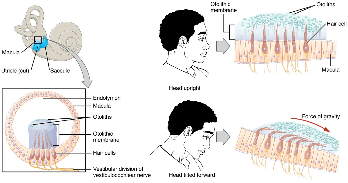 OtoconiaTiny calcium carbonate crystals that help the inner ear sense gravity and linear motion.OpenStax · Anatomy and Physiology · Fig. 14.11 · License can come loose from the
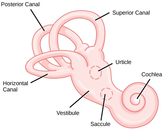 UtricleAn inner-ear organ that senses head tilt and linear motion. Otoconia can come loose from it and enter a semicircular canal.OpenStax · Biology · Fig. 36.15 · adapted from NIH · License and enter a semicircular canal. Moving debris can send a false signal of head rotation. Debris attached to the canal’s
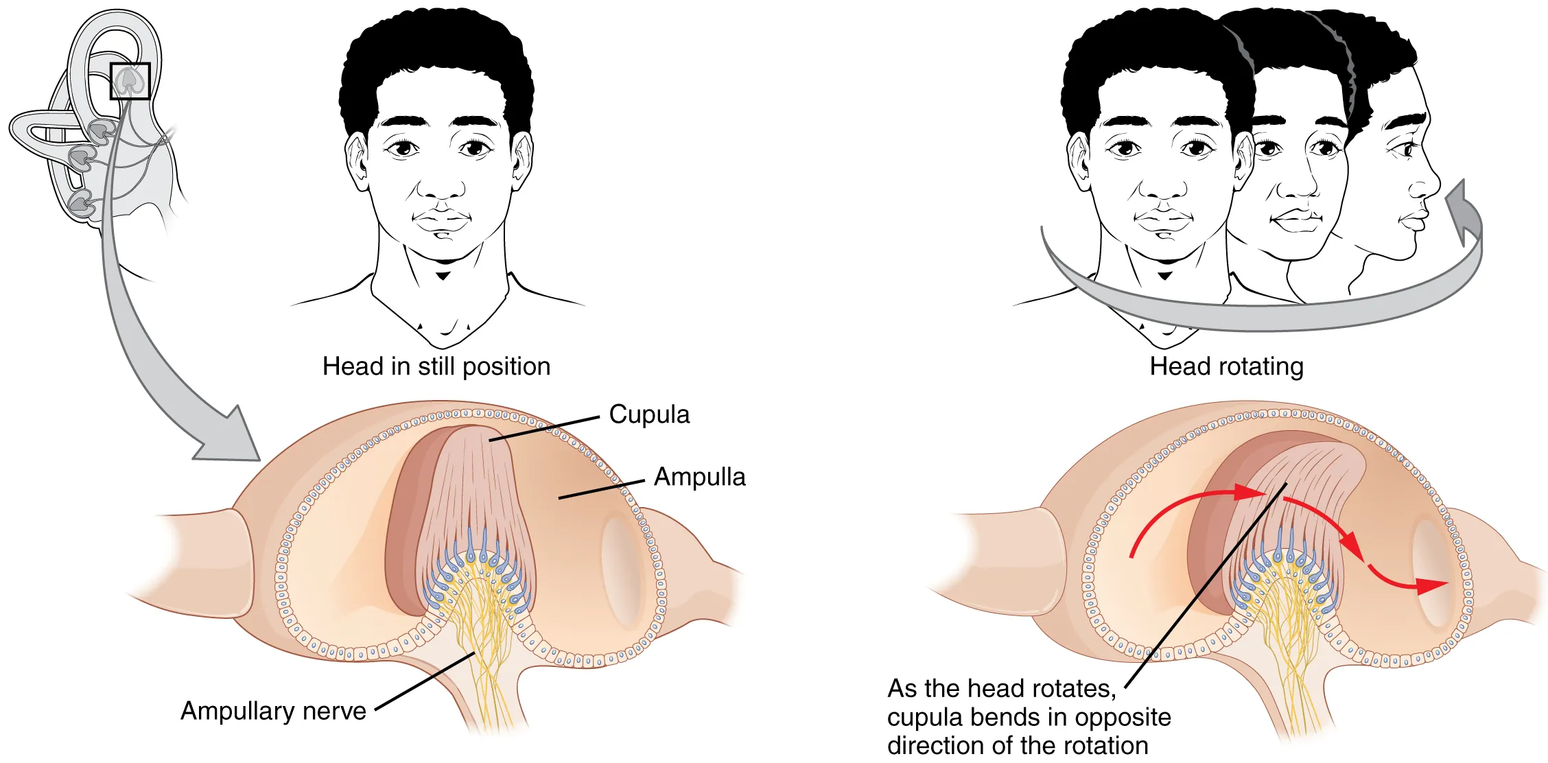 CupulaA flexible structure in the widened end of a semicircular canal. Fluid movement bends it and stimulates sensory cells.OpenStax · Anatomy and Physiology · Fig. 14.12 · License can cause a more sustained response while the position is held.
What do you look for during an exam?
Positional tests can reproduce vertigo and elicit an involuntary eye movement called nystagmus. Its direction and timing help you interpret which canal may be involved.
Start with the fast phase
The eyes drift slowly, then make a quick reset. Name the nystagmus by that quick movement. For torsion, watch the tops of the irises and describe the direction using the patient’s right and left.
Alexander’s law
With the head still, nystagmus that follows Alexander’s law gets stronger when the patient looks in the direction of the fast phase and weaker when they look the other way. The fast phase keeps the same direction. For example, right-beating nystagmus gets stronger on right gaze and weaker on left gaze.
Open Alexander’s law in the eye movement examples and switch between right and left gaze. Then open Direction-changing with gaze and switch sides again. In that example, the fast phase reverses.
From eye movements to exam findings
Start by asking what brought on the beats. Did the patient move only their eyes, or did their head change position relative to gravity? Then describe where the eyes snap, which position gives the stronger response, and how long the beats last.
With the head still: stronger beats or a reversal?
Ask the patient to look right and left while their head stays still. If the eyes beat rightward in both directions of gaze, with stronger beats on right gaze, that illustrates Alexander’s law. The direction stays the same as the intensity changes.
If the eyes beat rightward on right gaze and leftward on left gaze, the fast phase has reversed with gaze. Sustained reversal at moderate gaze angles can raise concern for a central gaze-holding problem. A few beats at the extreme edge of gaze can be normal.
See both examples in Read the eyes.
Right Dix–Hallpike: upward beats with a twist
After the patient reaches the right Dix–Hallpike position, there is a short pause. The eyes then snap upward toward the forehead while the tops of the irises twist toward the patient’s right ear, which is closer to the floor. The beats build and fade while the head stays in position.
What it suggests. This is the classic pattern for right posterior-canal BPPV due to canalithiasis, in which loose particles move and then settle.
Supine roll: use both sides to work out the likely ear
Compare right and left turns of similar size and speed. Say the fast phase beats right with the right ear down and left with the left ear down. Both beat toward the floor, so the pattern is geotropic. If it is clearly stronger with the right ear down, the right horizontal canal is more likely involved.
If the beats go toward the ceiling on both sides, the pattern is apogeotropic. For this form of horizontal-canal BPPV, the weaker side is usually the affected side. If the responses are similar, side assignment from intensity alone is uncertain.
While holding the position: when do the beats start and stop?
Latency is the delay from reaching the test position to the first beat. Duration is how long the beats continue while the position is held. In the brief posterior-canal example above, both the pause and the fading response fit free-moving particles.
If the same upbeating and torsional pattern continues beyond a minute, consider posterior-canal cupulolithiasis, where particles attached to the cupula can keep it deflected. The half Dix–Hallpike can help assess this possibility. Interpret the duration alongside the direction, position, and rest of the exam, since persistent positional nystagmus can have other causes.
Sources and page numbers
Bárány Society consensus on nystagmus (Eggers et al., 2019)
Choose the next step
Work through a sample case one finding at a time. After you answer, you’ll see the reasoning before the next finding appears.
Technique setup
Choose a test or maneuver to explore positioning, eye movements, and debris.
Canals and otoconia
Drag to inspect the canals. The table stays at the examiner’s angle. L and R mark the patient’s sides.