
BPPV Clinical Simulator
BPPV evaluation
Learn BPPV
Jump to a lesson
What is BPPV?
Benign paroxysmal positional vertigo (BPPV) typically causes brief episodes of dizziness or VertigoA false sense that you or your surroundings are moving. Patients may describe it as spinning, tilting, or simply dizziness. triggered by changes in head position relative to gravity. A patient might say, “The room moves when I roll over in bed,” or simply, “I get dizzy when I lie back.” Both descriptions are worth following up.
What to ask when a patient says “I get dizzy”
- What this answer can suggestLying back and rolling in bed can occur with either posterior- or horizontal-canal BPPV. A clear rolling trigger makes the supine roll test worth considering. Use the positional eye findings to identify the canal and side. Symptoms only on standing warrant an orthostatic assessment.. “Does it start when you lie back, roll over, look up, or stand up? If you roll in bed, does it happen on one side or both?”
- What this answer can suggestTime one episode. Brief movement sensations fit common forms of BPPV. Lingering nausea or unsteadiness can outlast an episode. Repeatedly prolonged movement sensations warrant a broader assessment. Use the duration of observed nystagmus when considering the debris mechanism.. “If you hold still in that position, how long does the main sensation of movement last? What lingers afterward?”
- What this answer can suggestMild unsteadiness can linger between BPPV episodes. New episodes that start without a position change, or dizziness that is continuous at rest, warrant a broader assessment.. “How do you feel before you move again? Does a new episode ever start while you’re already still?”
Bárány Society · BPPV diagnostic criteria and history notes, printed pp. 108–110
Brief, repeatable episodes that start with a position change support testing for BPPV when screening and positioning allow it. Dizziness that is already present at rest and worsens with movement calls for a broader assessment. Use the positional exam to identify the likely canal and side.
What happens in the inner ear?
Each inner ear has three Semicircular canalsThree fluid-filled ducts in each inner ear that sense head rotation in different planes.: horizontal, posterior, and anterior. They sense head rotation. Nearby, the UtricleAn inner-ear organ that senses head tilt and linear motion. Otoconia can come loose from it and enter a semicircular canal. helps sense head tilt and linear motion, such as when a car accelerates.
Tiny calcium carbonate particles called OtoconiaTiny calcium carbonate crystals that help the inner ear sense gravity and linear motion.OpenStax · Anatomy and Physiology · Fig. 14.11 · License Why otoconia may come loose Otoconia normally sit in a gel-like layer in the utricle. Degeneration of the crystals may make detachment more likely. BPPV can also follow a head injury. In many cases, there’s no identifiable trigger for the detachment. BPPV is more common in older adults and in women. It can also occur in younger adults, including after head injury. Human utricular otoconia study · Population study · AAO-HNSF guideline from the utricle and enter a semicircular canal. Inside a canal, those particles can send a false signal of head rotation.
CanalithiasisLoose otoconia move freely within a semicircular canal. means the particles move freely in the canal. In cupulolithiasisOtoconia adhere to the cupula, which can prolong the response while a position is held., particles cling to the 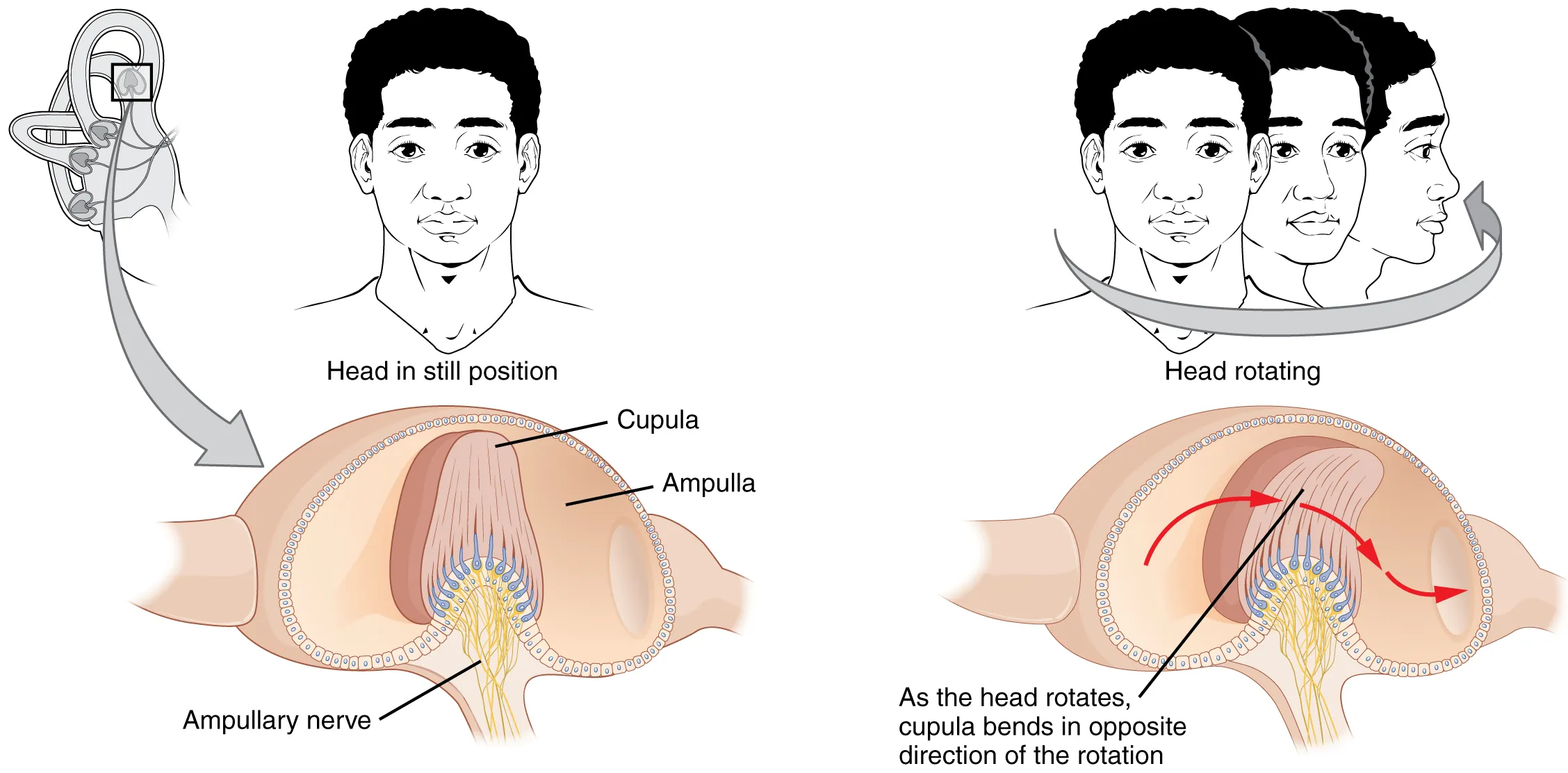 CupulaA flexible structure in the widened end of a semicircular canal. Fluid movement bends it and stimulates sensory cells.OpenStax · Anatomy and Physiology · Fig. 14.12 · License. Gravity can keep a weighted cupula bent while the head stays in the test position.
What do you look for during an exam?
Positional tests can reproduce vertigo and elicit an involuntary eye movement called nystagmusA repetitive, involuntary eye movement. Positional testing looks at its direction and timing.. Its direction and timing help you interpret which canal may be involved.
How a head turn becomes an eye movement
As the head starts to turn, fluid in the canals (endolymphThe fluid inside the semicircular ducts. Its movement relative to the duct bends the cupula.) initially lags behind. It bends the cupula, a flexible structure in the widened end of the canal, called the ampullaThe widened end of a semicircular duct that contains the cupula and sensory hair cells.. That bends the sensory hair bundles and changes how often the vestibular nerve fires.
Both ears send signals at rest. ExcitationAn increase in nerve firing above the resting rate. means firing increases above that baseline. InhibitionA decrease in nerve firing below the resting rate. means firing decreases. Matching canals across the two ears work as pairs. A rightward head turn excites the right horizontal canal and inhibits the left. The vestibulo-ocular reflex (VOR)A reflex that moves the eyes to compensate for head movement and helps stabilize gaze. moves the eyes left to help keep an object in view.
Watch the horizontal canals work together
Head still. Both canals fire at baseline.
Normal response to head rotation
Front view. The eyes counter-rotate during the head turn. The patient’s right is on your left.
Watch the pale cupula bend inside the gold ampulla. Its edges stay attached, and the dashed line marks its resting shape. The bend is exaggerated so you can see it.
Each tick is one nerve signal. Compare both rows over the same amount of time. The tick counts are illustrative.
What the patient feels. The brain uses the difference between the two canals’ firing rates as a cue that the head is turning. Here, that cue matches the actual turn.
This paired increase and decrease is called push–pullDuring rotation in the plane of a canal pair, firing increases on one side and decreases on the other.. During the head turn, one horizontal canal sends more signals while its partner sends fewer.
What changes when debris enters a canal?
The previous demo showed the normal reflex during an actual head turn. For BPPV, watch for positional nystagmus after the head reaches the test position. In this supine roll example, the examiner turns the head to the right, then holds it right ear down. With loose particles in the right horizontal canal, gravity can keep them moving after the head stops. That can drive a slow eye drift even while the head is still.
Right supine roll test
Face up. Choose a version of the same head turn.
What the patient may feel. Compare what happens after the head stops.
Face up · same view as the table
During a beat, the eyes drift slowly toward the patient’s left, then reset quickly to the right.
The white dot marks the nose in both head-end views. The highlighted canal is the right horizontal canal. Gold particles represent loose otoconia. The patient’s right is on your left in the eye close-up. Playback timing and motion are illustrative.
Watch the eyes drift toward the patient’s left, then snap back toward the right. When this cycle repeats, it’s called jerk nystagmusRepeated slow eye drift followed by a quick reset in the opposite direction.. The quick reset is the fast phaseThe quick reset in jerk nystagmus. Its direction gives the beat its name., so this example is called right-beatingThe quick reset goes toward the patient’s right. In a front view, it goes to your left..
For an upward or downward beat, watch the movement relative to the face. For torsionRotation of the eye around its line of sight. Describe where the top of the iris twists., watch the tops of the irises twist toward the patient’s right or left ear.
Before positional testing, watch the eyes with the head still
The roll-test example showed beats after the head changed position. In a clinical exam, first establish what the eyes are doing before you move the head. An oculomotor screen checks how the eyes hold gaze, line up, and follow or shift between targets. Findings at this stage can point to a need for a broader assessment before you interpret the positional response.
Start with straight-ahead gaze. With the head still, look for Spontaneous nystagmusBeats seen in straight-ahead gaze with the head still, without a provoking maneuver.. Note which way the eyes beat.
Then compare right and left gaze. Keep the head still and ask the patient to look to either side at a comfortable angle. Watch for Gaze-evoked nystagmusBeats brought out or changed by looking away from straight ahead while holding the head still. and compare the fast phase. With Alexander’s lawDirection-fixed nystagmus typically intensifies when looking in the fast-phase direction., right-beating nystagmus grows stronger on right gaze and weakens on left gaze. A sustained reversal at moderate gaze angles can raise concern for a central gaze-holding problem. Brief beats at extreme gaze can occur normally.
In the examples below, compare Alexander’s law and Direction-changing with gaze. On each side, ask whether the beats change strength or actually reverse.
How to check eye alignment and tracking
Alignment. Alternate the cover from one eye to the other while the patient looks at a target. Watch the newly uncovered eye for a vertical correction, which can indicate SkewA vertical difference in eye alignment. Alternating cover can reveal a vertical correction..
Tracking. Ask the patient to follow a slowly moving target to check Smooth pursuitThe continuous eye movement used to track a moving target.. Repeated catch-up movements can show that the eyes are falling behind.
Target changes. Ask the patient to look back and forth between two stationary targets to check SaccadesRapid eye movements that shift gaze from one target to another.. Watch for consistent overshooting or undershooting. Consider these findings alongside the history and the rest of the exam.
Choose a test and interpret the response
Before you position the patient
Use the history and the eye screen to decide whether positional testing fits. Check whether the patient can safely tolerate the neck motion, transfers, and head support. Use a suitable modification or seek help when positioning is limited. New focal neurologic signs, severe new headache or neck pain, or inability to sit or stand unaided warrant urgent assessment.
Choose the positional test
Use Dix–HallpikeA positional test used to look for a characteristic posterior-canal BPPV response. to check for posterior-canal BPPV. If the first side is negative, check the other side as tolerated. If the history still fits and Dix–Hallpike brings out horizontal beats or no nystagmus, use the supine roll testA test for horizontal-canal BPPV that compares eye movements with each ear turned down while the patient lies on their back. to check the horizontal canals. Vertigo without visible nystagmus leaves the canal and side uncertain.
Start by asking what brought on the beats. Did the patient move only their eyes, or did their head change position relative to gravity? Then describe where the eyes snap, which position gives the stronger response, and how long the beats last. A horizontal reversal between right- and left-ear-down roll positions can be expected in horizontal BPPV. A reversal when only gaze changes is a different finding.
Right Dix–Hallpike: upward beats with a twist
After the patient reaches the right Dix–Hallpike position, there is a short pause. The eyes then snap upward toward the forehead while the tops of the irises twist toward the patient’s right ear, which is closer to the floor. The beats build and fade while the head stays in position.
What it suggests. This is the classic pattern for right posterior-canal BPPV due to canalithiasis, in which loose particles move and then settle. Excitation of the right posterior canal drives a slow downward and twisting movement. The fast reset goes upward and twists toward the right ear.
Supine roll: use both sides to work out the likely ear
Compare right and left turns of similar size and speed. Say the fast phase beats right with the right ear down and left with the left ear down. Both beat toward the floor, so the pattern is geotropicThe fast phase beats toward the floor in both ear-down positions during the roll test.. If it is clearly stronger with the right ear down, the right horizontal canal is more likely involved. In the usual geotropic form, the right-ear-down turn moves fluid toward the ampulla of that canal and excites it. With the left ear down, the affected canal is inhibited and the beats are usually weaker.
If the beats go toward the ceiling on both sides, the pattern is apogeotropicThe fast phase beats away from the floor in both ear-down positions during the roll test.. For this form of horizontal-canal BPPV, the weaker side is usually the affected side. If the responses are similar, side assignment from intensity alone is uncertain.
While holding the position: when do the beats start and stop?
Clinically, ask whether the patient still feels movement after the head is held still, and how long it lasts. Record what they feel and what you see in the eyes separately. Use the eye pattern across test positions to help identify the canal and side.
LatencyThe time between reaching the test position and the onset of nystagmus. is the delay from reaching the test position to the first beat. DurationHow long the nystagmus continues while the test position is held. is how long the beats continue while the position is held. In the brief posterior-canal example above, both the pause and the fading response fit free-moving particles.
If the same upbeating and torsional pattern continues beyond a minute, consider posterior-canal cupulolithiasis, where particles attached to the cupula can keep it deflected. The half Dix–HallpikeA variation with the head supported slightly above supine to help evaluate a sustained posterior-canal response. can help assess this possibility. Interpret the duration alongside the direction, position, and rest of the exam, since persistent positional nystagmus can have other causes.
Downbeating on a positional test
Predominantly downward beats with a small twist can occur with rare anterior-canal BPPV. Downbeating positional nystagmus also raises concern for a central cause. Review the torsion, timing, and other exam findings before assigning a canal.
Ewald’s laws in the positional exam
The canal’s plane helps explain the direction of the eyes. The stronger response to excitation helps explain why the two sides of a roll test can produce different intensities.
Flow toward the ampulla (ampullopetalFlow toward the ampulla. This excites the horizontal canal.) excites the canal.
Flow away from the ampulla (ampullofugalFlow away from the ampulla. This excites the anterior and posterior canals.) excites the canal.
In the geotropic roll example, the stronger right-ear-down response supports right horizontal-canal involvement. The beat direction tells you the pattern. The difference in strength helps you work out the side.
Bárány Society · Canal excitation and slow eye movements, Fig. 3 · Experimental study · Horizontal canal response and Ewald’s second law
Sources and page numbers
Bárány Society consensus on nystagmus (Eggers et al., 2019)
- GRACE-3 · Acute dizziness by timing and triggers
- Alexander’s law · §2.1.1, printed page 69
- Gaze-evoked nystagmus · §2.2, printed page 75
- Posterior and horizontal canal patterns, including half Dix–Hallpike · §§2.3.1.1.1–2.3.1.1.2, printed page 77
- Anterior canal and central positional nystagmus · §§2.3.1.1.3 and 2.3.1.3, printed page 78
Match the maneuver to the findings
Before choosing a maneuver, name the likely canal, affected ear, and pattern. A repositioning maneuverA sequence of head and body positions used to guide displaced particles out of a canal. uses a sequence of positions to guide free-moving particles out of the canal. Positioning and head support affect which maneuver a patient can tolerate.
- Typical posterior-canal canalithiasis
- Use an Epley maneuverA repositioning sequence used for typical posterior-canal canalithiasis. on the affected side when appropriate. For example, brief upbeat and rightward torsional beats on right Dix–Hallpike support a right Epley.
- Geotropic horizontal-canal BPPV
- GufoniA side-lying maneuver used here for geotropic horizontal-canal BPPV. It starts on the unaffected side. or the barbecue rollA series of rolls around the body’s long axis used to reposition debris in horizontal-canal BPPV. are options when the affected side is clear. For geotropic Gufoni, the patient first lies onto the unaffected side. A right-sided diagnosis therefore starts with left side-lying.
- Apogeotropic horizontal pattern
- Check the weaker side and compare duration on both sides. Several mechanisms can produce this pattern. An apogeotropic maneuver such as AppianiA side-lying maneuver with an upward head turn used for some apogeotropic horizontal-canal presentations. may be appropriate when the findings and positioning support it. If the pattern converts to geotropic, repeat the roll test to guide the next choice.
- Unclear, sustained, or atypical findings
- Review the observed pattern and consider other causes before selecting treatment. If you cannot localize the canal or side, or the findings raise concern for a central cause, reassess or refer as appropriate.
Use the Technique library to review the setup and sequence for the test or maneuver you selected.
Reassess the response
Check whether the original trigger still brings on vertigo and whether the positional nystagmus has resolved, persisted, or changed direction. If a different pattern appears, localize it again before repeating a maneuver. If symptoms persist, evaluate for unresolved BPPV and other vestibular or central causes.
Discuss fall safety and the possibility of recurrence. Arrange reassessment within one month after treatment or observation to document the outcome.
Practice cases
Work through a patient’s history and exam findings one decision at a time. After each answer, you’ll see the clinical reasoning.
What would you do next?
Technique setup
Choose a test or maneuver to explore positioning, eye movements, and debris.
Canals and otoconia
Drag to inspect the canals. The table stays at the examiner’s angle. L and R mark the patient’s sides.